


Smoking is the most common cause of many chronic diseases, such as cancer, lung diseases, chronic bronchitis, stroke, emphysema, chronic obstructive pulmonary disease, and heart stroke. According to WHO, smoking tobacco kills around 8 million people each year, including smokers and non-smokers.
One of the main reasons for lung cancer is smoking. Second-hand smoke can also lead to lung cancer. The detection of lung cancer is tricky. Lung nodules form whenever there is illness or infection. Lung nodules are small clumps of cells also, known as granuloma. Granuloma is usually noncancerous lung nodules. But, when there is an uncontrollable growth of cells in the lung, these are called neoplasms. Malignant neoplasms are the cause of lung cancer.
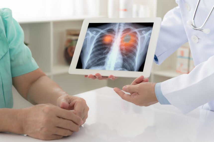
You must immediately consult a pulmonologist and get tests done for lung nodules, If you experience symptoms like coughing blood, wheezing, chest pain, chronic cough, hoarseness, and loss of appetite or weight loss.
A cancerous lung nodule occupies more and more space in the lung resulting in the symptoms mentioned above. If imaging tests results show that the nodule growth is faster, like in 25 days, you must go for further tests. There are chances of it being lung cancer.
More Read

Many tests are needed to detect lung cancer. A single test isn’t sufficient to find whether the lung nodule is cancerous or not. The diagnosis tests a pulmonologist will suggest you undergo can be invasive and non-invasive.
The diagnosis tests required for detection of lung cancer are as follows:
Basic tests:
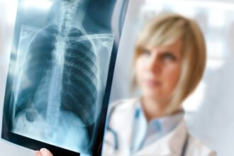
Chest X-Ray
Chest X-Ray is also known as chest radiograph. It uses a small amount of ionizing radiation to produce images inside the chest.
CT-Scan
Computed Tomography(CT) Scan is a non-invasive imaging technique. Radiographers control the X-Rays motion source to scan and produce required images.
CT-guided needle biopsy
In CT- guided needle biopsies, the radiologists use a needle to collect the tissue sample from the suspected tumor. It is an invasive technique.
Magnetic Resonance Imaging(MRI) Scan
In MRI Scan, radiologists use computer-generated radio waves and magnetic fields to generate images of tissues and organs in the body.
Positron Emission Tomography (PET) Scan
PET scans can detect abnormal activities more efficiently by producing detailed images of tissues and organs. It uses radiotracers to check metabolic changes. Doctors use this scan commonly to gain more cancer information.
Doctors use PET Scans often for the diagnosis of lung cancer in patients.
Major Tests for diagnosis:
For confirming that the patient has lung cancer, diagnosis of lung cells is a must. Tests for that purpose are:
Sputum cytology
A sputum sample is collected to test for cancer cells. Sputum is the mucus from the lungs while coughing. The sample usually is morning samples of three days back to back. This test helps find cancers starting in the major airways of the lungs.
Thoracentesis
A needle is inserted between the ribs to drain the fluid collected inside to check for cancer. Though sometimes, this fluid may be a result of an infection or heart failure.
Needle biopsy
In needle biopsies, a hollow needle collects a small sample from the suspicious area in the body. Needle biopsy doesn’t require a surgical incision. There are types of needle biopsy, fine-needle aspiration(FNA) biopsy, Transtracheal/transbronchial FNA, and core biopsies.
In FNA, the doctors use a thin hollow needle to withdraw aspirate cells. It is done to check cancer in lymph nodes.
During bronchoscopy or endobronchial ultrasound, a needle passes through the trachea or bronchi, it is known as transracial/transbronchial FNA.
Bronchoscopy
In bronchoscopy, the doctor uses a bronchoscope. A bronchoscope is a thin, flexible tube with a light and lens on end. A bronchoscope is put inside the body through natural openings like the nose or mouth down the throat into the trachea and airways of the lungs.
Nowadays, navigational bronchoscopy is more effective than traditional bronchoscopy. Navigational bronchoscopy has more benefits than traditional bronchoscopy.
Body Vision Medical is setting a new standard in lung cancer diagnosis with its Body Vision System. It uses artificial intelligence to produce real-time intraoperative CT images. It is compatible with any conventional C-arm available in the hospital.
Body Vision helps pulmonologists locate the exact area of the lesion and it provides visual tool-in-lesion confirmation. This image-guided biopsy approach is proven to maximize diagnostic yield. It seamlessly integrates with all existing tools and equipment in the bronchoscopy suite to provide superior clinical outcomes.
The Body Vision system has a portable main unit, a tablet for navigation control, and an optional procedural kit for those looking to use Body Vision as an end-to-end solution for navigational bronchoscopy.
Some remarkable benefits of Body Vision’s intraoperative imaging system are:
- True, real-time imaging of lesion and lesion location.
- The disposable catheter is compatible with existing endobronchial tools.
- Intraoperative CT imaging eliminates any issue of CT-to-body divergence
- It is proven to maximize diagnostic yield.
- It is cost-effective.
- It helps in the early detection of lung cancer.
- It is portable and small and has a compact design.


