


Thanks to advancements in dental technology, patients may now get treatment that is less invasive, less painful and takes less time than ever before. Problems with waiting, preparation (having to wait for the image to be processed), the surroundings, and other factors often arose when the older, more conventional X-ray methods were employed.
Therefore, dental X-rays and the work of dentists in general have been profoundly altered by the advent of digital radiography. In our talks with family dentist Dr. Babin we discovered that digital dental radiographs (digital X-rays) are increasingly being used by dentists today for enhanced detection, diagnosis, treatment, and monitoring of oral disorders and illnesses.
When it comes to capturing high-quality pictures of gums, teeth, as well as other oral structures and problems, digital radiography has replaced conventional photographic X-ray film with digital X-ray sensors.
There are three primary ways to capture digital dental images: directly, indirectly, and semi-indirectly. The direct approach involves the use of a digital sensor that is implanted inside your mouth to capture visual data. In the indirect method, dental X-rays are scanned from film and viewed digitally. Semi-indirect digital imaging employs a scanner and sensor to digitize dental X-rays.


How does digital radiography work?
One must be familiar with the operation of standard x-ray equipment before attempting to comprehend their inner workings. Electromagnetic radiation, such as X-rays, can penetrate flesh and bone but not water. Your dentist will be able to see any cavities you may have since your bones reflect more light than soft tissue.
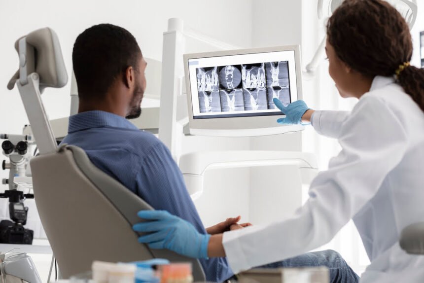
Dental x-rays taken digitally employ a sensor connected to a computer rather than film. To link this sensor to a computer, all you need is a bit of know-how and a cable. Digital radiography is a lot like old-school dental X-rays in terms of the procedure. With digital radiography, an intraoral sensor is used to take pictures of your teeth.
This is where traditional X-rays and their digital counterparts part ways. In reality, the digital option is an electronic gadget linked to a computer that only appears like the film used in conventional X-rays. You as well as your dentist may examine the photographs in real time, since the equipment takes snapshots and quickly displays them on a screen.
It is possible to capture intraoral and extraoral digital dental radiographs. The most frequent kind of dental X-ray, called an intraoral X-ray, has exceptional clarity and may be used to examine teeth and jaws for signs of decay, developmental issues, or disease.
In contrast to intraoral X-rays, which are used to pinpoint specific cavities, extraoral X-rays provide just a broad overview of the whole mouth. However, they are used in the diagnosis of impacted teeth, the tracking of jaw growth and development, and the detection of possible issues involving teeth, jaws, and other facial bones.
Dental imaging has several advantages
By using dental imaging, dentists can effectively identify and treat a wide range of gum and tooth issues. In order to examine oral health information, conventional techniques were previously employed. However, digital dental x-rays provide an alternative.
Some of the most significant advantages include the following:
- These digital sensors are used to collect the x-rays rather than the older technology of x-ray film, resulting in higher quality images. These sensors take high-resolution pictures that can be stored digitally. A dentist can make better decisions with more data at his disposal if the image is of high quality and accuracy. Images of the gums as well as other oral tissues may be recorded alongside those of the teeth for use in a computer database.
- The picture on conventional x-ray film can only be seen at its true size; however, there are options for enlarging the image. Because digital photos may be expanded without losing quality, the dentist can examine a more detailed view of any abnormalities.
- Traditional x-ray film needed a developing time before it could be seen, whereas digital x-rays may be viewed instantly. The imaged region may now be seen instantly thanks to digital technologies. Images may be seen on the computer monitor immediately after being taken.
- Accuracy of Diagnosis – Increased Access to high-quality images and other data improves diagnosis accuracy. Patients may feel at ease knowing that their diagnostic and treatment plan are tailored to their unique oral anatomy. Hidden cavities, bone infections, tumors, gum disease, and other abnormalities may not be obvious to the naked eye, but may be uncovered by digital x-rays.
- You may save both time and money by Minimizing the number of treatments needed is one of the main benefits of digital x-rays for early diagnosis. Thus, patients might just save both money and time by avoiding more intrusive procedures.
- Researchers have discovered a 70% reduction in radiation compared to conventional x-ray machines, which is a significant improvement. The potential for harm from x-ray exposure, both in the short and long term, may be mitigated by reducing the dose.
- The dentist clinic may preserve these records in electronic storage forever. Digital photographs may be stored safely in the cloud, eliminating the need for filing cabinets full of paper documents.
- You may request a hard copy of your digital x-rays if you’ll need to keep or show them to anybody else. In addition, the insurance claims procedure may be sped up by sending these pictures via computerized means.
- Digital x-rays may help reduce pollution in the environment since they do not need the use of chemicals to develop film. Capturing photos digitally eliminates the need for potentially dangerous chemical byproducts.
Special Education and Training
Digital radiographs may be taken by dentists, dental assistants, dental hygienists, and oral and maxillofacial radiologists who have received specialized training in this area. In most cases, state dental practice laws or dental board rules stipulate less strict radiographic training standards for dental office staff compared to those required for medical X-ray workers.
The availability and education in new equipment, supplies, and methods; infection control protocols; and continuous education in the safe and appropriate use of radiation devices are all factors to be considered when planning training.
Final words
Digital radiography reduces radiation exposure, but still you should use it only when required. Pregnant women and toddlers should wear thyroid collars and lead aprons.
Pregnant women may safely receive up to 4 radiographs each appointment visit, although most physicians and patients wait until the pregnancy is ended. Emergency X-rays for pregnant women are usually safe. “Double lead aprons” reduce radiography exposure to almost nothing. Breastfeeding and pregnancy do not delay X-rays.


