


Scientists at UCLA and at the California NanoSystems Institute have developed a small portable microscope that attaches to the camera module of a smartphone and detects nano particles including bacteria and viruses.
Scientists at UCLA and at the California NanoSystems Institute have developed a small portable microscope that attaches to the camera module of a smartphone and detects nano particles including bacteria and viruses.
The microscope device is light and portable and could be used in remote locations to monitor how well treatments for infections are working by detecting viruses and bacteria without the need for bulky and expensive microscopes and lab equipment. The device weighs less than half a pound.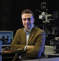

Aydogan Ozcan (image at right) and his colleagues developed the portable imaging system to address the need for a less expensive, powerful but compact microscope that could be uses in areas with limited resources.
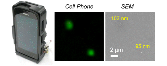
“This cellphone-based imaging platform could be used for specific and sensitive detection of sub-wavelength objects, including bacteria and viruses and therefore could enable the practice of nanotechnology and biomedical testing in field settings and even in remote and resource-limited environments,” Ozcan said. “These results also constitute the first time that single nanoparticles and viruses have been detected using a cellphone-based, field-portable imaging system.”
The microscope was used to detect polystyrene beads as small as 90-100 nanometers and this was validated using a scanning electron microscope (see photo above, courtesy ACS).
The microscope was also used to image individual HCMV particles. HCMV is a member of the herpes virus family and it can cause mortality in those whose immune system is compromised. It is also one of the leading causes of virus-associated birth defects such as mental retardation and deafness. A single HCMV particle is about 150-300 nanometers. (For comparison, a human hair is about 100,000 nanometers thick).
Ozcan is the principal investigator on the research. He is the founder of the mobile microanalysis startup company, Holomic LLC.
The scientists conclude:
“Given its high sensitivity and field-portability, our cellphone-based fluorescence imaging platform could be useful for specific imaging of various fluorescently labeled specimen such as bacteria and viruses in field settings. Therefore, it holds significant promise for various point-of-care applications such as viral load measurements or other biomedical tests conducted in remote or resource-limited environments.”
Sources:
http://www.acs.org/content/acs/en/pressroom/presspacs/2013/acs-presspac-september-25-2013/improved-smartphone-microscope-brings-single-virus-detection-to-.html
http://pubs.acs.org/doi/ipdf/10.1021/nn4037706
http://newsroom.ucla.edu/portal/ucla/ucla-researchers-smartphone-microscope-248254.aspx
To read other posts in this exclusive ongoing series, please visit the Mobile Health Around the Globe main page. And if you have a Mobile Health Around the Globe story to tell, please post a comment below or email me at joan@socialmediatoday.com Thanks!

