
Earlier Detection of Metastases
Stephen M. Bravo, MD, and his team at Sand Lake Imaging hope to help cure prostate cancer – or at least transform it from an acute and mortal threat into a manageable chronic disease. His team was therefore in no way astonished when Bravo suggested they participate in a clinical study aimed at earlier detection of lymph node metastases.
Detecting Abnormal Lymph Nodes
When looking at the background of the study, Dr. Bravo had to rely on size criteria such as whether a lymph node achieved the size of at least one centimeter in all dimensions. Lymph nodes smaller than one centimeter may also harbor disease, especially metastases from prostate cancer, but there was no unique tool to be able to detect this disease early on. And even when a lymph node reached one centimeter in size, it was uncertain as to whether that represented a myelodysplastic invasion from metastatic disease or whether it was just a reactive, normal inflammatory process that frequently occurs in the body, especially in reaction to prostatectomy or radiation therapy. Dr. Bravo states that their goal was to define early lymphatic disease before it became metastatic to a size greater than one centimeter. This requires both excellent imaging capabilities as well as efficient quantitative 3D visualization tools.
More Read

Precision Imaging for Prostate Cancer
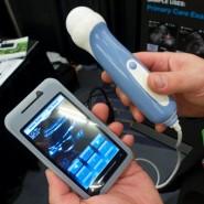
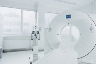
Dr. Bravo and his team are using Feraheme (Ferumoxytol) to detect metastatic disease in nodes as small as three millimeters. This drug accumulates in normal lymph nodes but is not absorbed in prostate cancer cells within the lymph nodes. Patients are given the contrast agent on their first day of imaging exams; they also have a computed tomography (CT) scan to localize the smallest of the lymph nodes. Feraheme takes 18 to 24 hours to show up as a hyper-intense lesion under magnetic resonance imaging (MRI). So, the actual MRI imaging is done on the second day using a 3-tesla system.
Multiple Imaging Modalities
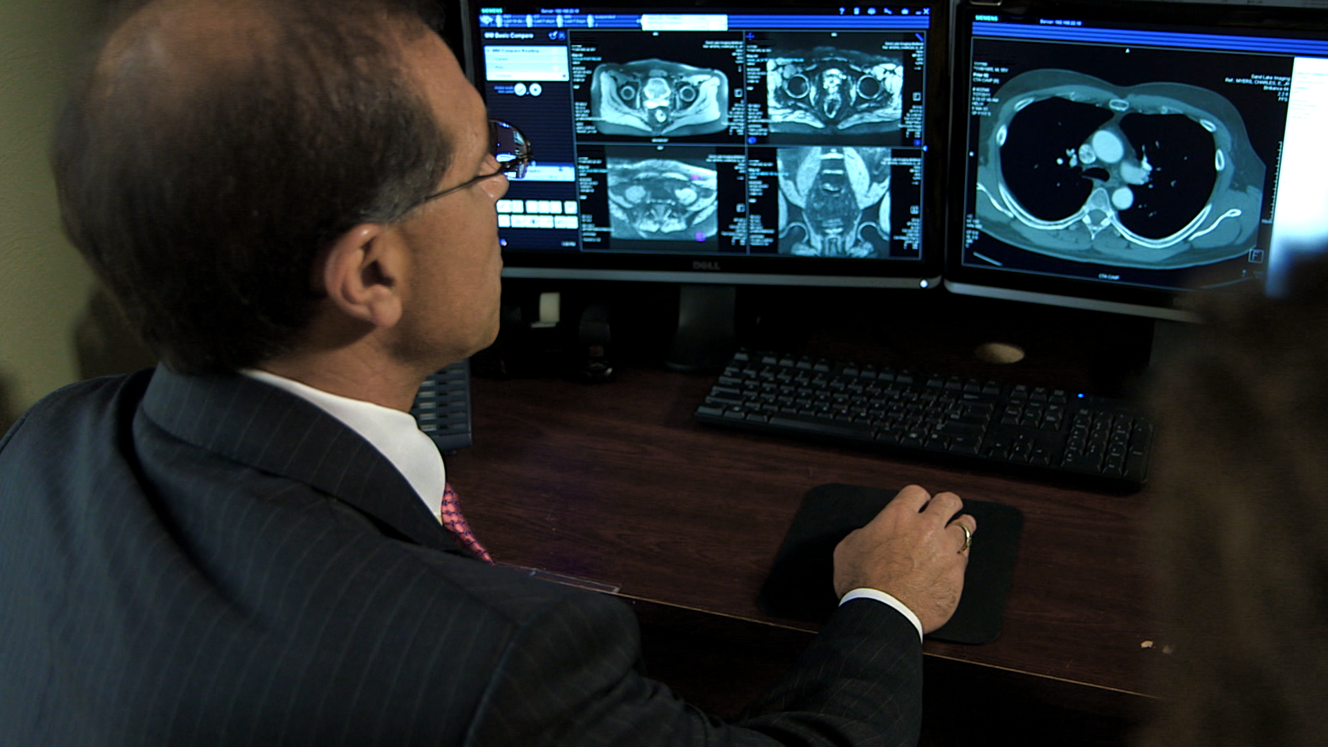
The CT images are then used to determine that the lymph nodes follow the appropriate signal characteristics on the MRI images – that is, that they all absorb the Feraheme contrast agent and show up accordingly on the MRI images. Positive lymph nodes that do not show up on the MRI scan as having absorbed the contrast agent are subsequently treated at affiliated clinics using precise radiation therapy. The next day, patients have a positron emission tomography/computed tomography (PET∙CT) scan with F18 Sodium Fluoride. PET∙CT is an important diagnostic tool for evaluating body metastatic disease and its accuracy in determining micro-metastatic disease has been proven in several studies for various cancers.
Multimodality Challenges
With three modalities and gigabytes of images, however, preparation, diagnosing, and reporting of images could have been complicated. According to Dr. Bravo, the biggest challenge for technologists in radiology is the constant pressure to be effective. syngo®.via3 helps with this challenge by making sure that the data sent over to the radiologists is right the first time, and is concise. One of the reasons why Sand Lake Imaging decided to go with the imaging software syngo.via was because they don’t have a 3D lab with staff processing images and sending them to the radiologists. Dr. Bravo felt that there is significant added value when combining the different modalities with syngo.via, enabling a far more precise and efficient workflow.
In conclusion, thanks to syngo.via, the team of Sand lake Imaging could not have carried out the Feraheme research.
Read more articles like this one on Medical Solutions Online – the Magazine for Healthcare Leadership. http://sie.ag/1bGqJIg
1 http://www.cancer.org/cancer/prostatecancer/detailedguide/prostate-cancer-key-statistics. Last accessed Oct 4th, 2013
2 The outcomes achieved by the Siemens customers described herein were achieved in the customer’s unique setting. Since there is no “typical” hospital and many variables exist (e.g. hospital size, case mix, level of IT adoption) there can be no guarantee that others will achieve the same results.
3 syngo.via can be used as a standalone device or together with a variety of syngo.via-based software options, which are medical devices in their own rights.