
Among the many trends and buzzwords floating around RSNA 2013, one of the key areas that seems to be popping up everywhere is “tomosynthesis,” which is 3D imaging using X-Ray technology.
Among the many trends and buzzwords floating around RSNA 2013, one of the key areas that seems to be popping up everywhere is “tomosynthesis,” which is 3D imaging using X-Ray technology. With enhancements being made to DT technologies, as well as numerous laws being written related to breast density, tomosynthesis is sure to be an important topic in the medical imaging community for a long time to come. Yesterday, Carestream presented “Stationary Chest Tomosynthesis System using Distributed CNT X-ray Source Array,” with the University of North Carolina School of Medicine. The results of this study showed the feasibility of a stationary chest tomosynthesis system. This has the ability to improve image quality and enhance detection of small lung nodules and other chest pathology.
In addition to our presentation, multiple sessions have focused on the benefits of tomosythesis. Two in particular that caught our attention were focused about digital breast tomosynthesis (DBT) and its superiority to conventional mammography in breast cancer detection, and a session focused on how tomosynthesis is more beneficial in detecting lung cancer. The former looked to expand upon the usual benefits of using DBT, which are reduced recall rates, improved diagnostic accuracy, and improved cancer detection. From there, Pragya A. Dang, M.D., of Massachusetts General Hospital, Boston, looked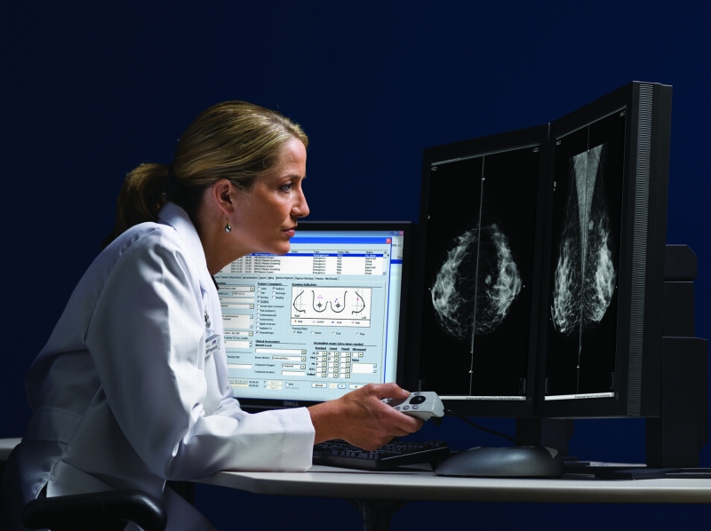
James T. Dobbins III, Ph.D., associate professor of radiology at Duke University, used dual-energy imaging and also looking at a broader range of expertise among radiologists when analyzing lung nodules. Dr. Dobbins saw that tomosynthesis had a threefold improvement in sensitivity, which is consistent with studies done in the past. He concluded that tomosynthesis is much better than conventional radiology when it comes to detecting lung nodules, and offered three options tomosynthesis implementation strategies:


- Using it as a problem-solving tool for suspicious findings on radiography
- Using it as alternative to CT for tracking changes in nodules over time, though Dr. Dobbins did state that additional studies on this would need to be conducted to validate this option
- Implementing it as a lower dose, lower cost model for lung cancer screening
From these studies, it is clear that we have only seen the beginning of DT and DBT. Study after study are showing the benefits of this technology, and as future studies are conducted, it becomes much more likely that we will start seeing wide-spread usage of tomoysnthesis for more accurate and efficienct diagnoses.

-150x150.jpg)