

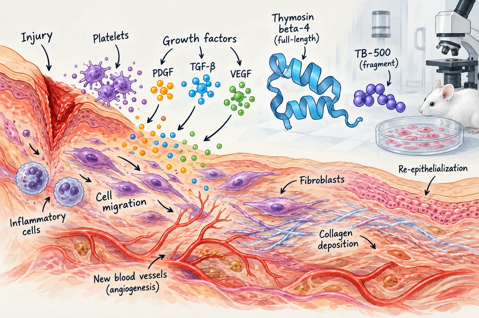
Injury triggers a lot of signals at once. Not one single switch. Platelets fire off growth factors, and – increasingly in experimental work – specific peptide fragments show up alongside them in the literature on wound closure, vascular regrowth, and post-injury recovery. To make sense of this field, you have to pull apart three things that tend to get mashed together: the biology of repair itself, the molecules researchers use to study it, and the wide gap between a promising cell-culture result and anything resembling a clinical outcome.
Researchers study tissue repair using cell cultures and animal injury models to examine growth-factor signaling, cell migration, and tissue changes. Interpreting those experiments depends on the molecule tested, the injury model, and the timing of measurements. Findings about full-length thymosin beta-4 do not automatically establish effects for synthetic fragments or clinical benefits in people.
Growth factors set the process in motion
Within minutes of damage, platelets dump out a mix of signaling proteins – PDGF, TGF-beta, VEGF, that crowd. These recruit inflammatory cells, kick fibroblasts into gear, and get new blood vessels forming. Researchers have picked apart this cascade for decades, using cell culture, rodent wound models, and more specialized setups like corneal injury or cardiac ischemia models. The sequence itself is pretty well nailed down at this point. What’s murkier is how to intervene in it usefully. That’s where the more targeted molecules come in.
No single peptide controls the entire healing process. Tissue repair depends on coordinated interactions between immune cells, blood vessels, fibroblasts, extracellular matrix components, growth factors, and numerous signaling molecules.
Sky Bloom IT in The Science of Healing Peptides: How Researchers Study Tissue Repair and RegenerationTiming complicates things further. VEGF showing up during new vessel formation isn’t doing the same job as TGF-beta signaling during later matrix remodeling. Researchers can measure both in a “wound healing” experiment and still be looking at two quite different events. That’s one reason broad claims about faster repair don’t tell you much on their own. You need to know what was measured, and when.
Actin dynamics and directed cell migration
Cell migration toward a wound depends heavily on the actin cytoskeleton – specifically, the push and pull between monomeric G-actin and filamentous F-actin. Thymosin beta-4, a 43-amino-acid protein sitting in most human cells, binds G-actin and keeps that balance in check. Back in 1997, a study in the FASEB Journal found that thymosin beta-4 stimulated directed migration of human umbilical vein endothelial cells – a result that shaped the angiogenesis research built around this protein ever since (Malinda, Goldstein & Kleinman, 1997). Two years later, a Journal of Investigative Dermatology study reported faster re-epithelialization and more collagen deposition in rat wound models treated with the same protein (Malinda, Sidhu & Mani, 1999).
Synthetic fragments as research tools
This part gets confusing fast, mostly because TB-500 and thymosin beta-4 are often treated as interchangeable names online. They aren’t quite that simple.
An analytical study examined material identified as TB-500 and found an N-terminally acetylated fragment corresponding to amino acids 17-23 of thymosin beta-4 – Ac-LKKTETQ. That matters when reading the research. The much larger literature on full-length thymosin beta-4 can’t automatically be used as evidence for the fragment.
More Read

For labs working specifically with this material, TB-500 peptide for sale belongs in the research-supply context. What matters experimentally is knowing exactly which molecule is being studied, instead of assuming results from the parent peptide carry over unchanged.
What the animal data shows
Most of what’s known about thymosin beta-4 and tissue repair comes from animal studies. Not human trials. In a mouse myocardial infarction model, the protein activated integrin-linked kinase, drove cardiac cell migration and survival, and reduced infarct-related damage (Bock-Marquette et al., Nature, 2004). Separately, applied topically, it sped up corneal re-epithelialization and cut down inflammatory cell infiltration after alkali injury in mice (Sosne et al., Experimental Eye Research, 2002).
Different injury models, similar story each time. That consistency is worth something. It still isn’t evidence of a clinical effect in people. Those are two different claims.
And the models themselves matter. A corneal injury study tells researchers something different from a skin-wound experiment; neither is a stand-in for cardiac tissue. Even when two studies point in roughly the same direction, the cells involved, route of administration, and outcome being measured may be completely different.
Why skin is a common tissue-repair model
Skin is experimentally accessible, which makes it one of the best organs for studying responses to tissue damage and repair. The review Wound repair and regeneration: Mechanisms, signaling, and translation describes how skin research has helped reveal fundamental principles in regenerative biology. That gives skin-wound experiments value beyond the particular wound being studied. For your team reading peptide studies, though, accessibility explains the choice of model; it does not remove the differences between skin, corneal, and cardiac injury described above.
Why the preclinical-to-clinical gap matters
A mechanism isn’t a treatment. Mouse and rat skin isn’t human skin – thickness, healing rate, immune response, all of it differs. The cardiac studies leaned on ischemia models that don’t capture how messy and variable a real human infarction actually is. Even a mechanism as well-documented as actin sequestration doesn’t carry over cleanly into a different species, or a different injury entirely.
Some early-phase human trials of the native protein have looked at things like chronic wound care. Results there are limited, and they shouldn’t be read as proof that anything works reliably. The synthetic research fragments modeled on thymosin beta-4? They haven’t gone through that process at all.
That’s an easy distinction to lose when the studies get summarized online. A cell result becomes an “effect,” an animal result gets described as a “benefit,” and before long the experimental model has disappeared from the story altogether. In this area especially, the model is part of the result.
Research use only
Peptides discussed here, including synthetic thymosin beta-4 fragments, are sold strictly for laboratory and in vitro research. Not for human or animal administration. Not for diagnosis, treatment, or disease prevention. Nothing in this article should be read as a recommendation for personal use.
Matching peptide materials to the experiments you compare
Comparing tissue-repair experiments requires matching the actual peptide and experimental conditions, because a shared product name does not establish equivalent material. For your laboratory, the relevant checks include molecular identity and purity documentation, alongside the cell model, assay design, and concentration used in each study.
A mislabeled peptide can compromise results before the assay begins. HealthLab Peptides’ discussion of experimental variables identifies material identity, purity, handling, and assay conditions as factors that can affect findings. When your team compares a TB-500 experiment with a thymosin beta-4 paper, check the documented molecular identity against the sequence studied. A full-length protein and an acetylated fragment remain different materials even when both appear under the same tissue-repair label.
Frequently Asked Questions
Can you buy peptides for research purposes?
To make sense of this field, you have to pull apart three things that tend to get mashed together: the biology of repair itself, the molecules researchers use to study it, and the wide gap between a promising cell-culture result and anything resembling a clinical outcome.
Sources
- Malinda KM, Goldstein AL, Kleinman HK. Thymosin beta 4 stimulates directional migration of human umbilical vein endothelial cells. FASEB Journal. 1997;11(6):474-481. DOI: 10.1096/fasebj.11.6.9194528.
- Malinda KM, Sidhu GS, Mani H, et al. Thymosin beta4 accelerates wound healing. Journal of Investigative Dermatology. 1999;113(3):364-368. DOI: 10.1046/j.jid.1999.00708.x.
- Philp D, Badamchian M, Scheremeta B, et al. Thymosin beta4 and a synthetic peptide containing its actin-binding domain promote dermal wound repair in db/db diabetic mice and in aged mice. Wound Repair and Regeneration. 2003;11(1):19-24.
- Bock-Marquette I, Saxena A, White MD, DiMaio JM, Srivastava D. Thymosin beta4 activates integrin-linked kinase and promotes cardiac cell migration, survival and cardiac repair. Nature. 2004;432(7016):466-472. DOI: 10.1038/nature03000.
- Sosne G, Szliter EA, Barrett R, Kernacki KA, Kleinman H, Hazlett LD. Thymosin beta 4 promotes corneal wound healing and decreases inflammation in vivo following alkali injury. Experimental Eye Research. 2002;74(2):293-299.
- Esposito S, Deventer K, Van Eenoo P. Synthesis and characterization of the N-terminal acetylated 17-23 fragment of thymosin beta 4 identified in TB-500, a product suspected to possess doping potential. Drug Testing and Analysis. 2012. DOI: 10.1002/dta.1402.
- Sosne G, Qiu P, Goldstein AL, Wheater M. Biological activities of thymosin β4 defined by active sites in short peptide sequences. FASEB Journal. 2010;24(7):2144-2151.